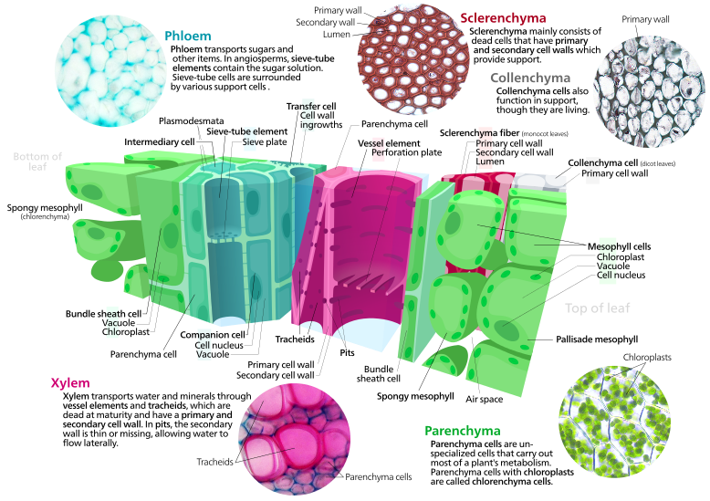
File:Plant cell types.svg

此SVG文件的PNG预览的大小:800 × 564像素。 其他分辨率:320 × 226像素 | 640 × 451像素 | 1,024 × 722像素 | 1,280 × 903像素 | 2,560 × 1,805像素 | 1,900 × 1,340像素。
原始文件 (SVG文件,尺寸为1,900 × 1,340像素,文件大小:7.16 MB)
文件历史
点击某个日期/时间查看对应时刻的文件。
| 日期/时间 | 缩略图 | 大小 | 用户 | 备注 | |
|---|---|---|---|---|---|
| 当前 | 2013年4月15日 (一) 23:11 |  | 1,900 × 1,340(7.16 MB) | IsadoraofIbiza | cut and moved text, added leaf side labels, photo labels |
| 2013年4月15日 (一) 00:03 |  | 2,000 × 1,360(7.24 MB) | IsadoraofIbiza | fixing clipped chloroplast | |
| 2013年4月14日 (日) 20:44 |  | 2,000 × 1,360(7.25 MB) | IsadoraofIbiza | center | |
| 2013年4月14日 (日) 20:40 |  | 2,000 × 1,360(7.25 MB) | IsadoraofIbiza | text tweak | |
| 2013年4月14日 (日) 20:31 |  | 2,000 × 1,360(7.25 MB) | IsadoraofIbiza | User created page with UploadWizard |
文件用途
以下2个页面使用本文件:
全域文件用途
以下其他wiki使用此文件:
- ar.wikipedia.org上的用途
- ast.wikipedia.org上的用途
- be-tarask.wikipedia.org上的用途
- bn.wikipedia.org上的用途
- bs.wikipedia.org上的用途
- crh.wikipedia.org上的用途
- cv.wikipedia.org上的用途
- en.wikipedia.org上的用途
- en.wikibooks.org上的用途
- es.wikipedia.org上的用途
- fr.wikipedia.org上的用途
- hu.wikipedia.org上的用途
- ja.wikipedia.org上的用途
- ka.wikipedia.org上的用途
- kn.wikisource.org上的用途
- ko.wikipedia.org上的用途
- krc.wikipedia.org上的用途
- la.wikipedia.org上的用途
- lbe.wikipedia.org上的用途
- os.wikipedia.org上的用途
- pt.wikipedia.org上的用途
- ru.wikipedia.org上的用途
- ru.wikinews.org上的用途
- sah.wikipedia.org上的用途
- simple.wikipedia.org上的用途
- sr.wikipedia.org上的用途
- ta.wikipedia.org上的用途
- uk.wikipedia.org上的用途
- xal.wikipedia.org上的用途
- zu.wikipedia.org上的用途


